CSF electrophoresis interpretation | OSCE station
Following is an OSCE station based on data interpretation. Data interpretation OSCE's are fairly straighforward and less time consuming
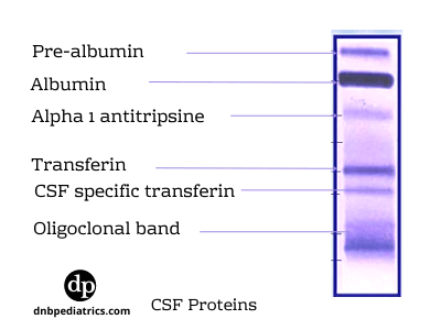
You are shown the following picture

Question1. Identify the test done on the CSF sample?
Answer
This is an image of CSF electrophoresis. Classically CSF agarose gel electrophoresis is used for this.
Question2. What abnormality does it show?
Answer
Oligoclonal bands
Question3. What is the differential diagnosis based on the abnormality shown?
Answer
See the anwer below in explanation
Question4. What other method is used to detect the abnormal finding?
Answer
Isoelectric focusing. It is most sensitive method for detection of the OCB.
CSF Electrophoresis
CSF electrophoresis is a qualitative test performed to identify different types of protein present in CSF.
It uses an electric charge to separate various proteins into a pattern of bands and the thickness of the bands depends on the concentration of that protein.
Normally a very little amount of protein is present in CSF, But when the blood-brain barrier is damaged due to various inflammatory conditions, say due to infective or autoimmune conditions, protein can leak into the CSF
Various proteins seen in CSF electrophoresis
Normal values of proteins in CSF
| Total protein | 15-45 mg/dL |
| Pre-albumin | 0.0-3.1 mg/dL (2%-7%) |
| Albumin | 8.4-34.2 mg/dL (56%-76%) |
| Alpha1 globulin | 0.0-3.1 mg/dL (2%-7%) |
| Alpha2 globulin | 0.0-5.4 mg/dL (4%-12%) |
| Beta globulin | 0.0-8.1 mg/dL (8%-18%) |
| Gamma globulin | 0.0-5.4 mg/dL (3%-12%) |
| Oligoclonal bands | None |
| IgG | 0.0-4.5 mg/dL |
Useful formulas
IgG Index
Reference range, 0.34 to 0.7. Higher levels simply mean higher production of IgG in CSF. Higher levels point toward autoimmune disorders, SSPE, and chronic infections.
Albumin Index
Reference range, 0 to 9. The sole producer of albumin is liver. Higher levels in CSF means a damaged blood-brain barrier.
What is an oligoclonal band?
Oligoclonal bands are bands of immunoglobulin that are seen on protein electrophoresis and indicate inflammation of the CNS, they are non-specific. Refer to image 1
They can be seen both in CSF and plasma (paired samples). More than two bands should be seen in the CSF sample.
They can be seen in up to 90% of patients with multiple sclerosis (MS).
Differential Diagnosis
Oligoclonal bands are also found in
- Multiple sclerosis, (commonly seen)
- Devic's disease or neuromyelitis optica
- Systemic lupus erythematosus,
- Neurosarcoidosis,
- SSPE
- Subarachnoid hemorrhage,
- CNS Syphilis,
- CNS Lymphoma
- ADEM
Author

Ajay Agade | DNB(Pediatrics), FNB(Pediatric Intensive Care), Fellowship in Pediatric pulmonology and LTV
Ajay is a Paediatric Intensivist, currently working in Pediatric Pulmonology & LTV at Great Ormond Street Hospital NHS, London