Pediatric ECG | 12 Leads and ECG Paper
1. Understanding ECG paper
ECG paper is a standardized special graph paper and has the following peculiar features.
Understanding ECG paper horizontally
- Each 1 mm/small) horizontal box is equal to 0.04 second (40 ms),
- 5 Small boxes form 1 large box marked by darker pink lines. It represents 0.20 sec 20 milisec intervals. so,
Every single small box horizontaly = 0.04 msec
Every single large box = 0.2ms
Understanding ECG paper vertically
- Vertically, the ECG graph measures a given wave’s height called amplitude or deflection. The standard calibration is 10 mm that is 10 small boxes which is equal to 1 mV of deflection.
Every single small box = 0.1 mV
Every single large box = 0.5 mV - On occasion, mainly when the waveforms are very small, the standardised calibration can be doubled. Now 1 mv equals 20 mm. When the waveforms are very large, half standard may be used 5 mm equals one mv. For example, in Pericardial effusion and ventricular hypertrophy respectively.
Speed of ECG paper
- The ECG waves are recorded on a special paper divided into one mm grid-like boxes.
- The ECG paper runs at a default speed 25 mm/sec i.e. 25 small boxes per second.
- The paper speed can be increased to 50 mm/secto define waveforms better.
Paper speed and voltage are usually printed on the bottom of the ECG for reference and are very important to interpret accordingly, since these parameters can be changed when the situation demands.
OSCE tip - While attempting observed OSCE station on reading ECG, convey that you have confirmed name, DOB, reg. no. as well as speed and calibration. This will give you more perks.

2. 12 lead ECG system
12 lead ECG combines six limb leads and six chest leads to capture the electrical activity of the heart from 12 different viewpoints.
Each lead depicts whether the electrical impulses in the heart are flowing towards that lead or away from it. The electrical current flowing towards a lead generates a positive deflection on the ECG graph, whereas the current flowing away causes a downward negative deflection.
What are Unipolar and Bipolar leads in ECG?
Unipolar leads are depicted to the voltage between a single positive electrode and a 'central' point of reference produced from the other electrodes.
Bipolar leads record the graph by measuring the voltage between two electrodes. For example, lead I show the voltage between the left arm and the right arm electrode.
There are 3 bipolar (I, II, and III), 3 augmented limbs (aVR, aVL, and aVF) and 6 unipolar or precordial or chest leads (V1–V6)
Bipolar ECG leads
- Lead I - RA act as the negative pole and LA as the positive pole.
- Lead II - RA act as the negative pole and LL as the positive pole.
- Lead III - LA as the negative pole and LL as the positive pole.
- II = I + III - This is known as Einthoven’s law (related to voltage)
- Lead aVR - LA+LL as the negative pole and RA as the positive pole.
- Lead aVL - RA+LL as the negative pole and LA as the positive pole.
- Lead aVF - RA+LA as the negative pole and LL as the positive pole
Unipolar leads
- V1, V2 nears RV and gives us data of Anterior-posterior forces
- V3, V4- near the septal wall and
- V5 and V6 near the LV, gives us the left to right forces.
Overall, they give information on all parts of the heart except its posterior surface.
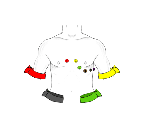
How to place ECG leads in Children
Electrodes must be positioned in accordance with AHA recommendations. Lead Positioning is the key to avoiding misinterpretation since these are not uncommon errors.
Start attaching Red, yellow, Green, and Black (ride your green bicycle) leads in a clockwise direction starting from the right wrist
Bipolar lead placement
- Red electrode – right forearm, proximal to the wrist
- Yellow electrode – left forearm, proximal to the wrist
- Green electrode – left lower leg, proximal to ankle.
- Black electrode – right lower leg, proximal to the ankle
|
| How to place ECG lead - note that the limb leads are usualy placed near wrist and ankles |
Unipolar lead placement
- V1 - 4th intercostal space, right sternal edge
- V2 - 4th intercostal space, left sternal edge.
- V3 Midway in between V2 and V4
- V4 5th intercostal space, mid- clavicular line
- V5 Left anterior axillary line, same horizontal level as V4
- V6 Left mid-axillary line, same horizontal level as V4 and V5
One of the OSCE station can be demonstrating lead placement on a given dumy patient
What is an observed OSCE station? All OSCE postsWatch this space for next post - What is noise/interference in ECG trace and how to troubleshoot."
Image attribution - Baklazan99 CC BY-SA 4.0 via Wikimedia Commons
Further reading
Author

Parvez Patel | DCH, DNB (Pediatrics), FNB (Pediatric Cardiology), Advanced Fellow RACP
Parvez has completed Pediatric residency from LTMC Mumbai and is currently working as advance fellow in cardiology in Australia
💡 Join the Discussion!
🩺 Help us refine this article — share corrections or additional information below. Let's elevate the accuracy of knowledge together! 💉💬